


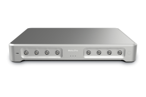
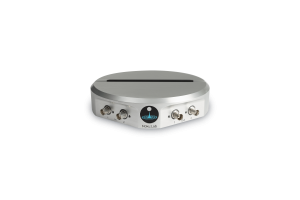
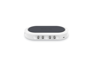

Quantum microscopy is a subfield of quantum optics that leverages the principles of quantum mechanics, namely two-photon entanglement, to achieve resolutions and sensitivities in imaging with the potential to exceed those of classical microscopy techniques. In a significant advancement in the field of quantum microscopy, researchers from the University of Bristol in the United Kingdom have developed a technique that allows for sub-micrometer precision in depth imaging. This new method, detailed in their latest study published in Physical Review A, utilizes entangled two-color photons in a Hong-Ou-Mandel (HOM) spectroscopy setup. To accelerate their research, the scientists are using Moku:Go, an FPGA-based device that delivers a reconfigurable suite of test and measurement instruments. Leveraging the Data Logger and Oscilloscope instruments, the team has developed a novel way to leverage quantum mechanics to image samples with high depth resolution.
The challenge
Traditional imaging techniques, such as electron beam microscopy or optical microscopy, often require high levels of illumination to achieve optimal resolution. However, high power can generate a number of problems, including nonlinear behavior in materials, damage to sensitive biological tissue, and photobleaching. This is especially problematic when looking at live cells or other in situ samples, so a gentler method must be used, ideally one that can achieve the same level of precision as traditional imaging. The group of Dr. Jonathan Matthews at Bristol is looking to develop such a technique harnessing quantum technology, one which can achieve similar levels of precision but at lower power consumption.
The solution
The team’s innovation lies in their use of HOM microscopy for imaging, which leverages quantum interference. In this configuration, pairs of entangled photons are first created. These pairs of photons are sent along different optical paths, and then pass through a beam splitter before arriving at two different photodetectors. Information about the difference in optical paths can be deduced from the relative arrival times of the photon pairs by monitoring the intensity output of the interferometer. This setup allows one of the paths to function as a transmission microscope, with information about the sample depth being probed and carried by one of the photons in the pair. The group also adjusted the wavelength separation of these entangled photons by heating the crystal used to produce them, creating two-color photon pairs and allowing them to find the optimal resolution for their microscope. However, as seen in Figure 1a, the optical setup required to implement such a scheme is complex. A 404 nm laser beam is first sent through a polarizing beam splitter to split it into two paths. These two beams are used to pump a crystal (ppKTP) from two separate directions, which emits photon pairs as a result. These photons are then entangled by passing them through another partial beam splitter, and are finally coupled into single-mode fibers.To keep the photon-pair generation path phase-locked, a 750 nm CW laser passes in reverse through the setup to a separate pair of photodetectors. The output from these photodetectors is monitored with the Moku:Go Data Logger, which can detect a failure of the phase locking during the course of an experiment. Dr. Cyril Torre, a researcher in the lab of Dr. Jonathan Mathews from the University of Bristol group, says Moku:Go was a tremendous asset in his research.“We used the [Moku] Oscilloscope to make sure that the system was locked to the correct frequency,” he said. “We chose Moku:Go because we didn’t need super specs — we needed a multi-tool. It’s very user-friendly.” After generation, the entangled photon pairs pass to the microscopy setup, as shown in Figure 1b. One optical path transmits through the sample before recombination with its partner, using the HOM technique described above. A series of photodetectors then analyze the results.

Figure 1: The experimental setup used in the experiments. (a) Photon-pair generation, using a 404 nm laser in conjunction with a ppKTP crystal and polarizing beam splitter. The resulting entangled pairs pass through outputs A and B. (b) The HOM microscope. One beam path passes through the sample while the other directly enters the PBS. The resulting pairs are collected by an array of photodetectors.[1]
The result
The group demonstrated the efficacy of two-color HOM microscopy by imaging a semitransparent sample with varying depth features. The group performed a raster scan across the surface of the sample, subdividing it into 4,000 pixels and probing the depth at each one. The result, alongside a conventional optical microscope image, is shown in Figure 2. The precision of the measurement using the HOM setup was estimated to be around 1 μm, although this figure can be adjusted by tuning the frequency difference between the photon pairs. The group’s approach significantly reduces the illumination intensity required for imaging, reaching the performance of super-resolution spectroscopy with a probe intensity of only 10-8 W/cm2, which is 8 to 12 orders of magnitude lower than the power required to achieve such precision with classical techniques.

Figure 2: A comparison of two-color HOM measurement versus classical imaging. Left: 3D reconstruction of the sample using the depth calculated at each pixel. The estimated axial resolution is around 1 μm. Right: A classical optical microscope image of the same sample.[1] Dr. Torre and his colleagues have developed an optical imaging system that harnesses the power of two-color entangled photon pairs. The result is a HOM microscope with sub-micrometer precision, low illumination requirements, and a variable dynamic range, which presents a substantial benefit for the study of photosensitive biological samples and materials. Looking ahead, Dr. Torre sees space for reconfigurable solutions like Moku to become commonplace.“We also have one Moku:Pro and two Moku:Lab devices that are currently being used in other experiments in our group,” he said. “They’re working very well. We like the Moku software and the regular updates.”
Questions?
Get answers to FAQs in our Knowledge Base
If you have a question about a device feature or instrument function, check out our extensive Knowledge Base to find the answers you’re looking for. You can also quickly see popular articles and refine your search by product or topic.
Join our User Forum to stay connected
Want to request a new feature? Have a support tip to share? From use case examples to new feature announcements and more, the User Forum is your one-stop shop for product updates, as well as connection to Liquid Instruments and our global user community.
References
[1] C. Torre, A. McMillan, J. Monroy-Ruz, and J. C. F. Matthews. Phys. Rev. A. 108, 023726 (2023).