

Please update your browser. For the best Liquid Instruments experience, update to the latest version of Chrome, Edge, Firefox, or Safari. Update Microsoft Edge.
Liquid Instruments Logo
Products
Hardware Platforms
Moku:Delta
Moku:Delta
New
For advanced design and validation
Moku:Pro
Moku:ProFor versatile test and measurement
Moku:Lab
Moku:LabFor experimental research labs
Moku:Go
Moku:GoFor quick testing and debugging
Compare Hardware
Compare HardwareView a side-by-side of all Moku hardware
Technologies
GenInst Studio
New
Generate personalized instruments
Integrated InstrumentsProfessional-grade instruments at your fingertips
Neural NetworkFPGA-powered neural network integration
Multi-Instrument ModeCombine instruments to create a customized test system
Moku CompileCode, compile, and deploy to your Moku's FPGA
Software
MokuOSDownload the Moku app for Windows, macOS, iPadOS & visionOS
APIsPython, MATLAB, LabVIEW
UtilitiesCommand line and graphical tools for file conversion
Instruments
Analysis
Oscilloscope
Spectrum Analyzer
Frequency Response Analyzer
Lock-in Amplifier
Phasemeter
Logic Analyzer / Pattern Generator
Time & Frequency Analyzer
Data Logger
Generation
Waveform Generator
Arbitrary Waveform Generator
Control & Conditioning
PID Controller
Digital Filter Box
FIR Filter Builder
Laser Lock Box
Custom & Utility
GenInst Studio
New
Gigabit Streamer
Custom Instrument
Neural Network
Multi-Instrument Mode
Applications
Optics and Photonics
Microscopy and Spectroscopy
Photon Counting
Quantum Optics
Aerospace and defense
A&D Instrumentation
Component Test
Automated Testing
Semiconductor Test
Control Systems and Bode Analysis
MEMS Testing
AI and Machine Learning
Generative Instrumentation
New
Neural Networks
Education
Education and Research
Resources
App Notes
Blog
Case Studies
Coursework
Guides
Product Documentation
Publications
Webinars
White Papers
Support
Company
Contact Us
About Us
Leadership
Partners
News & Updates
Press
Events Calendar
Careers
Store
Moku:Delta
Moku:Pro
Moku:Go
Upgrade Moku:Pro
Upgrade Moku:Lab
Upgrade Moku:Go
0
Get a quote Buy Now
Build personalized instruments with GenInst Studio in minutes, not months. | Register for the webinar
Case studies

Simplifying two-color stimulated Raman scattering (SRS) microscopy

University of Washington researchers used Moku:Pro to perform a variety of experiments and extract the low-intensity SRS signals with one device

April 25, 2022

    Introduction

    At the University of Washington, researchers working on chemical imaging tools for applications like early cancer detection and understanding neurodegenerative disease progression use two-color stimulated Raman scattering (SRS) microscopy. Experiment setups typically include multiple, complex, high-performance instruments for real-time two-color SRS imaging or simultaneous imaging of two widely spaced Raman transitions. Using the Moku:Pro Lock-In Amplifier and Multi-Instrument Mode, they are now able to perform a variety of experiments and extract the low-intensity SRS signals with one compact, multi-channel device.

    The challenge

    SRS is a coherent Raman scattering process that allows for chemical imaging with both spectral and spatial information. In a typical setup it uses two synchronized pulsed lasers, namely pump and Stokes (Figure 1), to coherently excite the vibration of molecules. To extract the very small SRS signal from a noisy background, a high-frequency modulation and phase-sensitive detection scheme is necessary.
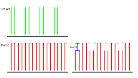
    Figure 1: The amplitude modulation transfer of Stokes to the pump beam due to SRS is detected. The demonstrated pump beam has a repetition rate of 80 MHz, and the Stokes beam has the same 80 MHz repetition but is also modulated at 20 MHz. The Δpump is extracted in this detection scheme.

    To perform experiments with real-time two-color SRS imaging, researchers must apply orthogonal modulation and detect both in-phase and quadrature signal components.
    “In most SRS microscopy experiments, the spectral range is limited to about 300 cm-1 because of limitations in the total bandwidth of the lasers,” said Dr. Dan Fu, Assistant Professor of Chemistry at the University of Washington. “One approach to circumvent this is to scan through wavelengths with a tunable laser, but that is slow and often insufficient for experiments that are time sensitive such as live cell imaging.”

    To overcome these limitations, researchers at the University of Washington use a third laser beam to enable simultaneous imaging of two widely spaced spectral regions, for example one in the fingerprint region (e.g. ~1600 cm-1 for Amide vibration) and one in C-H region (e.g. ~2900 cm-1 for protein), but this increases experiment setup footprint and complexity.

    Figure 2: HeLa cell SRS images taken with the Moku:Pro Multi-Instrument Mode setup at widely spaced apart Raman transitions.

    Read our ebook: Multi-channel lock-in detection in SRS microscopy and spectroscopy experiments

    The solution

    A quality lock-in amplifier is a critical hardware component in SRS microscopy experiments with a modulation transfer detection scheme. Moku:Pro’s Lock-in Amplifier provides an intuitive, precise, and robust solution for self-heterodyne signal detection in SRS microscopy experiments. The user interface allows for intuitive and powerful controls for extracting the low-intensity SRS signal.
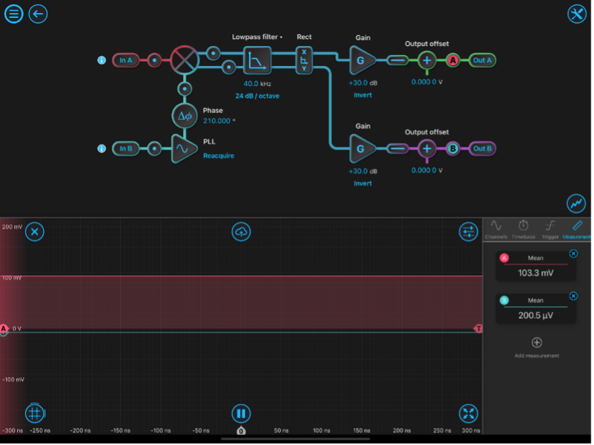
    Figure 3: Moku:Pro Lock-in Amplifier with typical single channel configuration settings.

    The Moku:Pro Lock-in Amplifier is configured with phase shift, low-pass filter and gain settings optimized for the experiment. The built-in probe points are used for real-time monitoring as setting are adjusted. Both X and Y outputs are made available for dual-channel imaging.

    In the case of three lasers, Moku:Pro Multi-Instrument Mode can be configured with two lock-in amplifiers, simplifying the system down to one device without compromises. This allows researchers to take two SRS images of large wave number difference simultaneously, utilizing one Moku:Pro to process two photodiode detector signals.

    Figure 4: Moku:Pro’s Multi-Instrument Mode with multi-channel lock-in amplifier configuration.

    The Multi-Instrument Mode configuration with two lock-in amplifiers for simultaneous SRS microscopy experiments is demonstrated in Figure 4. For the Lock-in Amplifier in Slot 1, input In 1 is the detected signal of the first photodiode, In 2 is the reference, Out 1 is the signal sent to an external data acquisition card, and Out 3 is discarded. For the Lock-in Amplifier in Slot 2, In 3 is the detected signal of the second photodiode, In 2 is once again the reference, Out 2 is the signal sent to external data acquisition card, and Out 4 is discarded. Each detected signal (Out 1 and Out 2) is maximized by adjusting their individual phase shifts before being sent to the data acquisition card. Slot 3 and 4 in this example are configured with Oscilloscopes but can be replaced with another Moku:Pro instrument. 
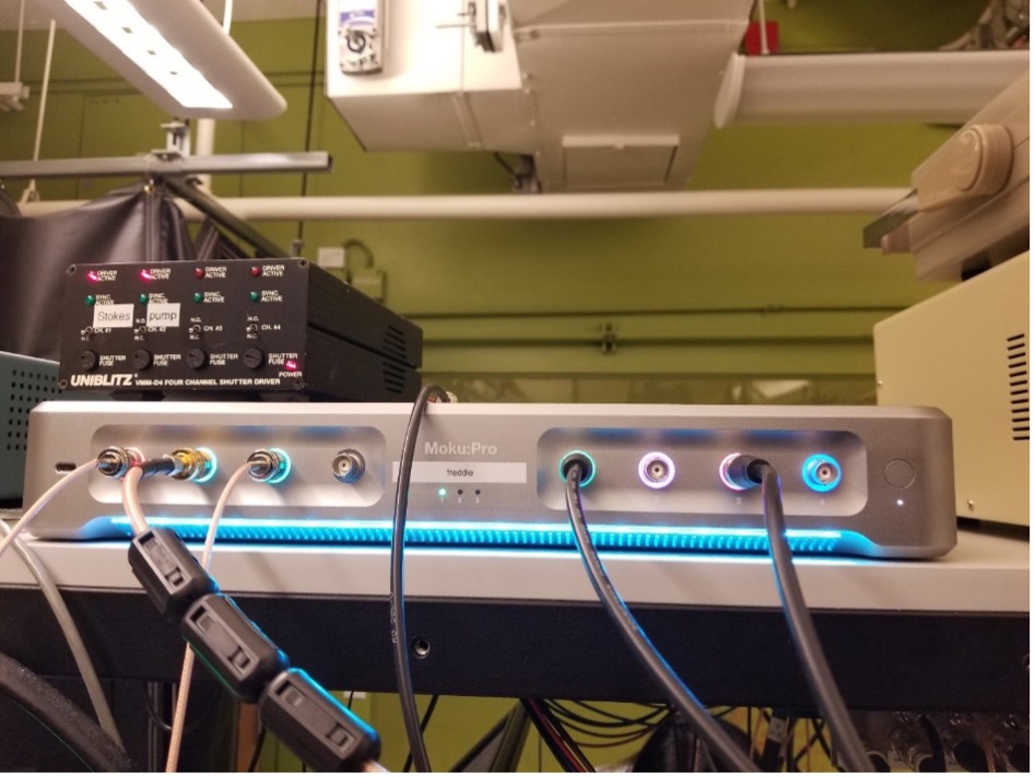
    Figure 5: Moku:Pro in Multi-Instrument Mode configured with two lock-in amplifiers with three input channels and two output channels in use.

    The result

    Moku:Pro’s Lock-In Amplifier provides an excellent solution for a multitude of SRS microscopy experiments. “The user interface allows for intuitive and powerful controls for extracting the low-intensity SRS signal and Moku:Pro’s Multi-Instrument Mode allows for complex imaging experiments on a compact system,” said Dr. Fu. From typical single channel SRS imaging, to dual-channel imaging, and even multi-instrument imaging, researchers at the University of Washington were able to simplify their experiment setup without compromises.

    Try Moku in demo mode Download the Moku app → Get answers to FAQs Visit Knowledge Base → Connect with Moku users Join the user forum →

    Other Recommended Case studies

    Case studies
    Phase Stabilization for Quantum Key Distribution with Moku:Pro

    Learn how researchers at the University of Hamburg are using Moku to improve signal fidelity for continuous-variable QKD.

    Featuring: Moku:Pro, PID Controller, Lock-in Amplifier, Laser Lock Box

    Date: Mar 16, 2026

    Case studies
    Enabling Dual-Wavelength Nanosecond Fiber Lasers for Multi-Contrast Photoacoustic Imaging with Moku:Pro

    Learn how researchers at the University of Hong Kong used Moku:Pro to generate synchronized shortwave-infrared nanosecond pulses and stabilize modulation depth for dual-wavelength photoacoustic imaging.

    Featuring: Moku:Pro, Waveform Generator, PID Controller

    Date: Dec-19-2025

    Case studies
    Teaching lock-in detection with Moku:Go

    Learn how students at IIT Madras are gaining hands-on experience in using the Lock-in Amplifier instrument on Moku:Go

    Featuring: Moku:Go, Lock-in Amplifier

    Date: Dec-15-2025

    Back to all Case studies

    Nav

    • Products
    • Instruments
    • Resources
    • Support
    • Company
    • Contact Us
    • Privacy Policy
    • Terms of Use

    Offices

    United States
    (619) 332-6230
    12526 High Bluff Dr.
    Suite 150
    San Diego, CA 92130

    Australia
    +61 2 6171 9730
    243 Northbourne Avenue
    Suite 1, Level 1
    Lyneham, ACT 2602

    Australia
    +61 3 7073 3594
    Suite 5C, Level 5,
    700 Swanston Street,
    Carlton, VIC – 3058


    Follow us

    LinkedIn YouTube X Facebook Instagram

    官方微信

    © 2026 Liquid Instruments. All rights reserved.